Eye and orbit
Innermost layer: the retina (nervous tunic)
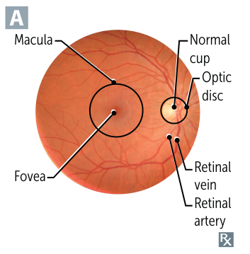
- Optic disc
- Formed by axons of ganglion cells that leave the eye to form the optic nerve
- Located medially to the fovea centralis
- Insensitive to light due to lack of photoreceptors (physiologic scotoma, i.e., blind spot)
- Contains the optic cup
- Central, cup-like depression in the optic disc
- Site of transversing of retinal vessels
- Point of exit for ganglion cell axons leaving the eye
- Macula
- An oval-shaped yellow spot on the lateral side of the optic disc, near the center of the posterior wall of the retina
- The very center (fovea) is avascular, the surrounding macular area have a blood supply from retinal artery branches
- Contains the fovea centralis
- A central depression in the macula (foveola)
- Contains the highest density of cones, each of which is connected to a single ganglion cell
- Point of sharpest vision (100% visual acuity)
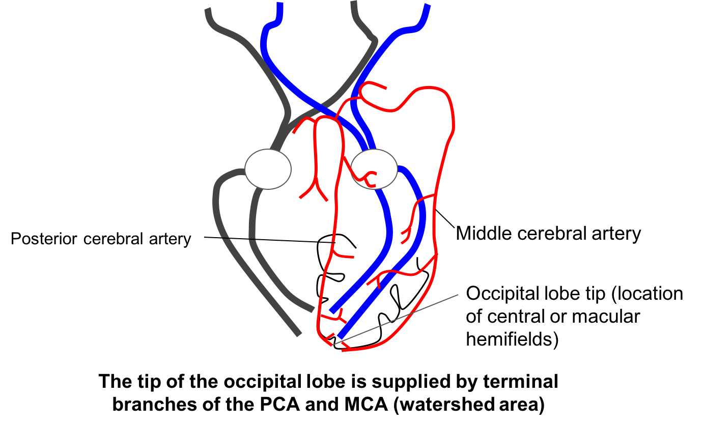
- Macular sparing: the macular region of the visual cortex has a dual blood supply. It receives blood from both the posterior cerebral artery (PCA) and the middle cerebral artery (MCA).
Nerves
-
Cranial Nerve III (Oculomotor)
- Function:
- Motor: Innervates Superior Rectus, Inferior Rectus, Medial Rectus, Inferior Oblique muscles (moves eye up, down, medially), and Levator Palpebrae Superioris (lifts eyelid).
- Parasympathetic: (Fibers travel on the outside) Innervates Sphincter Pupillae (pupil constriction) & Ciliary muscle (accommodation).
- Presentation (CN III Palsy): Eye is "down and out." Ptosis (eyelid droop) and a fixed, dilated pupil ("blown pupil").
- Key Associations:
- Compressive Lesion (e.g., PComm Aneurysm, uncal herniation): Affects superficial parasympathetic fibers first → mydriasis is an early sign. "Surgical" or "Painful CN III Palsy."
- Ischemic Lesion (e.g., DM neuropathy): Affects deeper motor fibers, sparing parasympathetics → "Pupil-sparing CN III Palsy."
- Function:
-
Cranial Nerve IV (Trochlear)
- Function: Innervates Superior Oblique muscle (SO), which causes intorsion and depression of the adducted eye. (Mnemonic: SO4)
- Presentation (CN IV Palsy): Vertical diplopia (worse when looking down, e.g., reading/walking downstairs). Head tilt away from the side of the lesion to compensate. Patients struggle looking down and in.
- Key Associations: Most common cause is head trauma (long, thin nerve). Can also be congenital or ischemic.
-
Cranial Nerve VI (Abducens)
- Function: Innervates the Lateral Rectus muscle (LR), which abducts the eye. (Mnemonic: LR6)
- Presentation (CN VI Palsy): Horizontal diplopia, worse on gaze toward the affected side. Inability to abduct the eye (esotropia - eye deviates medially at rest).
- Key Associations: Vulnerable to stretching from ↑ ICP (false localizing sign). Also common in DM, trauma, and pontine strokes.
-
Cranial Nerve V₁ (Ophthalmic Division of Trigeminal)
- Function: General sensation to the cornea, forehead, and upper eyelid.
- Clinical Relevance: Afferent limb of the corneal reflex. (Touching cornea → V₁ sensory input → bilateral blink mediated by CN VII).
- Presentation: Loss of corneal sensation (risk for ulceration), loss of afferent corneal reflex.
-
Cranial Nerve II (Optic)
- Function: Vision; afferent limb of the pupillary light reflex.
- Clinical Relevance: Lesion results in an afferent pupillary defect (APD) or Marcus Gunn pupil (paradoxical pupillary dilation in affected eye with swinging-flashlight test). Associated w/ optic neuritis (MS).
-
Autonomic Pathways
- Sympathetic: Dilation (mydriasis). Pathway: Hypothalamus → spinal cord (C8-T2) → superior cervical ganglion → travels along carotid artery to the eye.
- Horner's Syndrome: Lesion anywhere along this path → Ptosis (mild), Miosis (constricted pupil), Anhidrosis (decreased sweating). (Mnemonic: PAM is Horny). Associated w/ Pancoast tumor, carotid dissection.
- Parasympathetic: Constriction (miosis) via CN III (see above). Pathway: Edinger-Westphal nucleus → CN III → ciliary ganglion → short ciliary nerves.
- Sympathetic: Dilation (mydriasis). Pathway: Hypothalamus → spinal cord (C8-T2) → superior cervical ganglion → travels along carotid artery to the eye.